
May Thurner Syndrome | Iliac Vein Treatment Austin
Iliac Vein Compression Syndrome
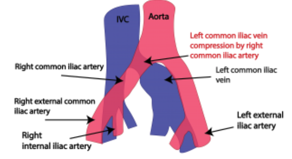
May Thurner Syndrome is a well-described anatomical abnormality involving the pelvic veins. This circulatory disorder most typically involves the left iliac vein that drains the left leg and left pelvic region. The left common iliac vein is the most commonly involved vessel. There are similar type of vein conditions that can occur in the right pelvis and leg as well, but it affects the left side of the body most frequently.
Anatomy of May Thurner Syndrome
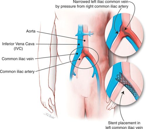
In May Thurner Syndrome there is found to be an abnormal compression of the left iliac vein between the right common iliac artery and the 5th lumbar vertebrae of the spine. The artery lays on top of the vein and the spine lays below the vein, so that the vein is almost “sandwhiched” by these structures. Although all individuals have their left iliac vein located adjacent to the iliac artery and spine, most individuals have enough space between these structures so that no significant compression of the iliac vein occurs. When not enough space exists between these structures, the iliac vein becomes abnormally compressed. Since the iliac vein is the softest and most compressible of the 3 structure (compared to bone and artery which are firmer / stiffer), it is the vein that becomes narrowed when the described anatomical condition is present.
It is theorized that chronic pulsation of the right iliac artery due to blood flow with each beat of the heart causes progressive chronic scar tissue to develop inside the vein wall and lumen of the iliac vein. This scar is referred to as intimal hypertrophy or intimal hyperplasia. When the abnormal veins are examined anatomically and under the microscope, a web or band of abnormal scar tissue known as fibrosis is seen. This scar tissue causes abnormal narrowing of the vein that is referred to as vein stenosis. This narrowing causes abnormal pressure elevation to develop within the leg veins and pelvic veins. Progressive obstruction of the flow of blood occurs.
How did May Thurner Syndrome Get Its Name
The term May Thurner Syndrome was given to this condition in 1957 by 2 scientists (May and Thurner) who discovered the anatomy in a large human autopsy series. In looking earlier in the medical literature, it was first described by McMurrich in 1908. It is felt to be a congenital issue of anatomy rather than a disease process caused by some other condition. It is not felt to be caused by trauma or infection.
Later in 1965, Crockett and Thomas coined the phrase “iliac vein compression syndrome” to better describe the condition. They called attention to the clinical symptoms that patients with May Thurner Syndrome present with, including leg pain and swelling. They also reported the long-term consequences of chronic venous insufficiency that results following thrombosis complications from May Thurner Syndrome. As a result, some medical literature (particularly in Europe) refers to May Thurner Syndrome as “Crockett’s Syndrome.”
Incidence and Gender
May Thurner Syndrome occurs in 25-30% of the population, although symptoms and complications from the anatomical condition only occurs in a small number of these individuals. Many patients remain asymptomatic throughout their lives. This condition is more frequently seen in females and it is theorized that prior pregnancies can increase the incidence of symptoms and complications from the condition. The most common age that individual present with the syndrome is between their 20s and 40s, although it can manifest at any age.
Deep Venous Thrombosis from May Thurner Syndrome
2-3% of all lower extremity and pelvic deep vein thrombosis (DVTs) are felt to occur secondary to May Thurner Syndrome. This explains why iliofemoral vein DVT is five times more likely to occur on the left side of the body. DVT symptoms include leg pain and leg swelling. Back and pelvic pain can also occur. Skin discoloration and dermatitis of the leg and foot can occur. If the blood clot travels it can float to the lungs, leading to chest pain, shortness of breath, and even sudden death from cardiac arrest. This movement of the clot is referred to as pulmonary embolism.
More delayed symptoms can occur in the legs as well, including heaviness and fatigue, chronic dermatitis, skin ulcers or wounds of the ankles and feet, leg asymmetry, and chronic leg pain or swelling.
Other May Thurner Symptoms
Other lower extremity symptoms can occur in addition to blood clots and Post-Thrombotic Syndrome, which are the most serious complications of the condition. These symptoms most frequently occur on the left side. They include swelling of the ankle or leg, discoloration of the ankle skin (usually tan, brown, or red), dermatitis of the ankle, foot, or leg, slow healing ulcers or wounds of the ankle or foot, and chronic leg pain. Other symptoms that can occur in the affected extremity include burning, itching, numbness, tingling, easy fatigue, heaviness, cramping, aching, and throbbing. Venous claudication is another well described symptoms.
When these symptoms occur, they are the result of elevated venous pressure inside the leg. Venous blood is trying to return from the leg to the heart, but is having difficulty doing so because of the anatomical resistance that is occurring in the pelvis vein. The pressure build up is from resistance ad obstruction at the level of the left common iliac vein. This pressure build-up tends to worsen at the condition progresses. Valve failure inside other leg veins can then occur over time, making the symptoms get worse. If a blood clot occurs due to May Thurner compression, then the symptoms can suddenly worsen.
May Thurner Syndrome Treatment
Please consult with a board-certified vascular surgeon before undergoing any therapy for May Thurner Sydrome. Treatment options will vary depending on the severity of the condition. Patients who develop blood clots are treated differently than those who do not. Some of the most common therapies are described below.
Stenting and Angioplasty
Putting a stent the left iliac vein is a common procedure performed for May Thurner Syndrome. This procedure is often advocated in cases involving a documented episode of Deep Venous Thrombosis (DVT) or other significantly symptomatic cases. This can be done following treatment of a blood clot or before a blood clot occurs. Stretching out the narrowing in the common iliac vein using a balloon, referred to as angioplasty, is often performed before or after the stent is inserted. Both stent and angioplasty are minimally invasive procedures that are performed through a catheter. The catheter is placed through a small puncture in the skin at the groin or behind the knee. Ultrasound and fluoroscopy x-ray is utilized in order to avoid any surgical incisions. No general anesthesia is needed, just local anesthetic. This is an outpatient procedure, with most patients being able to go home the same day. The success rate of the procedure is very high. Seek a consult with a board-certified vascular surgeon to determine if this is an option for you.
Medications For May Thurner Syndrome
Medications for treatment of May Thurner Syndrome are typically medications that diminish the tendency for your blood to form clots. Options can include Plavix, Aspirin, Coumadin / Warfarin, Xarelto, Pradaxa, Eliquis, Lovenox, Heparin, among other possibilities. Some patients do not require any medications. Medications recommendations will vary depending on your individual condition. Consults with a board-certified vascular surgeon to obtain recommendations.
Surgery for May Thurner Syndrome
Surgery is occasionally required for severe cases. Surgical options can include vein bypass procedures or venous reconstruction with an open pelvic surgery.
Thrombolysis for May Thurner Syndrome
Thrombolysis is a interventional procedure performed at the hospital. Thrombolysis is reserved for cases of May Thurner Syndrome that have resulted in blood clots known as deep venous thrombosis. During this procedure a medication called tPA (tissue plasminogen activator) is infused through a intravenous catheter and into the blood clot. The tPA medication works through a mechanism of actions to dissolve the blood clot. So thrombolysis is literally a clot busting therapy. A vein stent is usually required after the venous thrombolysis has been completed.
Venograms & IVUS
There are many forms of diagnostic imaging that are performed during the workup and treatment of May Thurner Syndrome. Venogram vein imaging is one critical compotent. Venogram involves injection of dye through an IV and into the vein to obtain detailed images of the problem. Venogram can be done with a CT scan (CT venogram or CTV), an MRI machine (MR venogram or MRV), or though more invasive catheterization procedures (catheter venography).
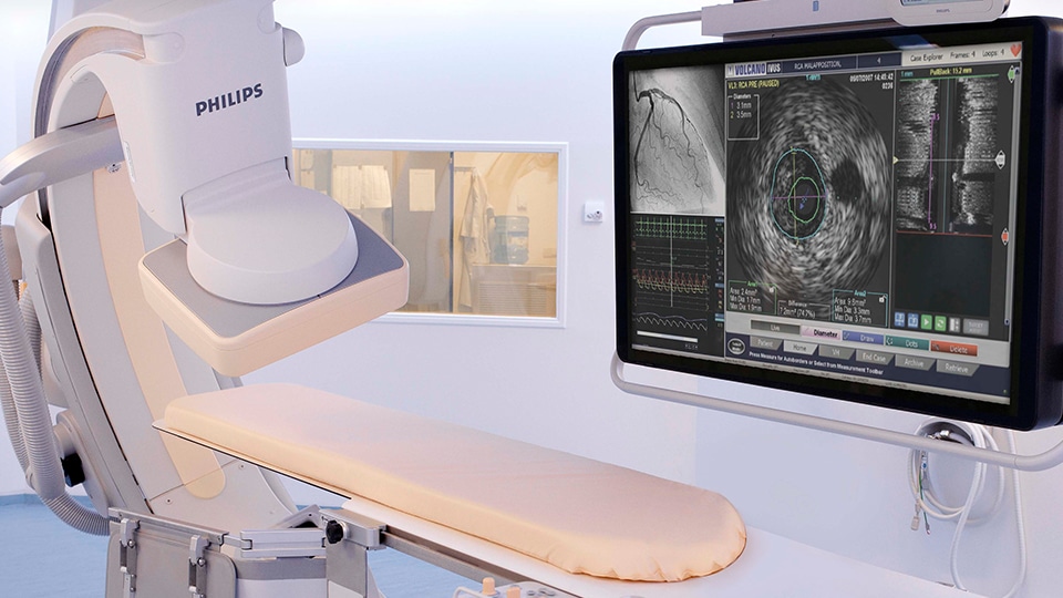
Even additional anatomical detail in May Thurner Syndrome can be achieved through the use of IVUS. IVUS stands for “IntraVascuar UltraSound.” During IVUS, a small fiberoptic catheter than contains a very this ultrasound transducer at its tip is introduced into the leg vein and guided up into the pelvic veins. Since the probe is inside the lumen of the vein where the blood is flowing, amazing detail of the iliac vein anatomy can be projected onto a ultrasound monitor for the physician to see.
May Thurner Syndrome Experts
At Austin Vascular Specialists, we are experts in the diagnosis and treatment of May Thurner Syndrome. We offer all of the latest state-of-the-art treatments available. Our board-certified vascular specialists has many years of experience treating May Thurner Syndrome. Contact us for a consultation.